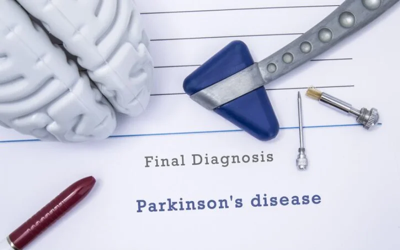
For years, Parkinson’s disease has been widely thought to stem from toxic iron buildup in the brain. But a growing body of evidence now suggests the opposite may be true: neurons affected by Parkinson’s could be starving for usable iron, even when total iron levels appear high.
The emerging idea, known as functional iron deficiency, proposes that while iron accumulates in certain brain regions most notably the substantia nigra it may be locked away in forms that cells cannot use. As a result, neurons may lack the biologically active iron required for dopamine production and energy metabolism.
This shift in thinking follows unexpected results from recent clinical trials. Studies testing the iron-chelating drug deferiprone, designed to remove excess iron from the brain, found that patients’ symptoms often worsened, particularly in early-stage Parkinson’s patients not yet receiving dopamine-based treatments. If iron overload were the main driver of the disease, researchers say, removing iron should have helped not harmed patients.
Rethinking iron’s role in Parkinson’s
The link between Parkinson’s disease and iron dates back decades. Imaging and post-mortem studies consistently show elevated iron signals in the substantia nigra, the brain region where dopamine-producing neurons degenerate. These findings fueled theories that excess iron causes oxidative stress and triggers iron-dependent cell death pathways such as ferroptosis.
But iron biology is more complex than once believed. Iron exists in multiple forms, and not all are biologically useful. Ferrous iron (Fe²⁺) is essential for enzymes involved in dopamine synthesis and mitochondrial function, while ferric iron (Fe³⁺) is largely inert and stored in proteins like ferritin or neuromelanin.
Crucially, MRI scans cannot distinguish between these forms. Researchers now argue that the “iron overload” seen in Parkinson’s may reflect sequestration of unusable Fe³⁺ rather than an excess of functional iron inside vulnerable neurons.
Clues from dopamine biology and early trials
The new perspective also revisits overlooked findings from the early days of Parkinson’s treatment. The disease’s hallmark motor symptoms arise from dopamine loss, and dopamine synthesis depends on tyrosine hydroxylase, an iron-dependent enzyme.
Biochemical studies in the 1970s and 1980s showed that iron strongly boosts this enzyme’s activity. Small clinical studies from that era even reported symptom improvement in Parkinson’s patients given iron supplements, with some able to reduce their dopamine medications. While these trials lacked modern controls, they hinted that iron deficiency not toxicity might impair dopamine production.
Supporting evidence from genetics and epidemiology
Additional support comes from rare genetic disorders known as neurodegeneration with brain iron accumulation (NBIA), where iron builds up in the brain but neurons nonetheless show impaired iron use and dopamine dysfunction. Animal studies further show that blocking iron uptake in dopamine neurons leads to cell death and Parkinson-like symptoms.
Population studies also suggest a link: anemia and recent blood donation have been associated with a higher risk of developing Parkinson’s, though researchers caution that these findings do not prove causation.
Implications for treatment
Together, these findings challenge the long-held belief that excess iron is a primary cause of Parkinson’s disease. Instead, they point to a paradox in which iron is present but biologically inaccessible—effectively starving neurons of a critical resource.
If confirmed, the implications could be profound. Rather than removing iron from the brain, future therapies might aim to restore iron availability, improve iron trafficking within neurons, or prevent iron from being locked away in unusable forms. Researchers stress that any such approach would need to carefully consider disease stage and existing dopamine treatments.
While more clinical trials are needed, the new model offers a compelling explanation for why iron-chelating therapies have failed—and opens a new chapter in understanding one of neurology’s most challenging diseases.